Kidney Stones Chest X Ray

Urolithiasis Radiology Reference Article Radiopaedia Org

Renal Calculus Pre And Post Eswl Radiology Case Radiopaedia Org

Xray Image Of Plan Kub Shows Kidney Stones Stock Photo Picture And Royalty Free Image Image 39887442

Plain Kidney Ureter Bladder Bilateral Kidney Stones And Stomach Download Scientific Diagram

Kidney Stones Collection Of Plain X Rays

Plain Kidney Ureter Bladder Bilateral Kidney Stones And Stomach Download Scientific Diagram
Cystine stones appear similar to other renal stones.
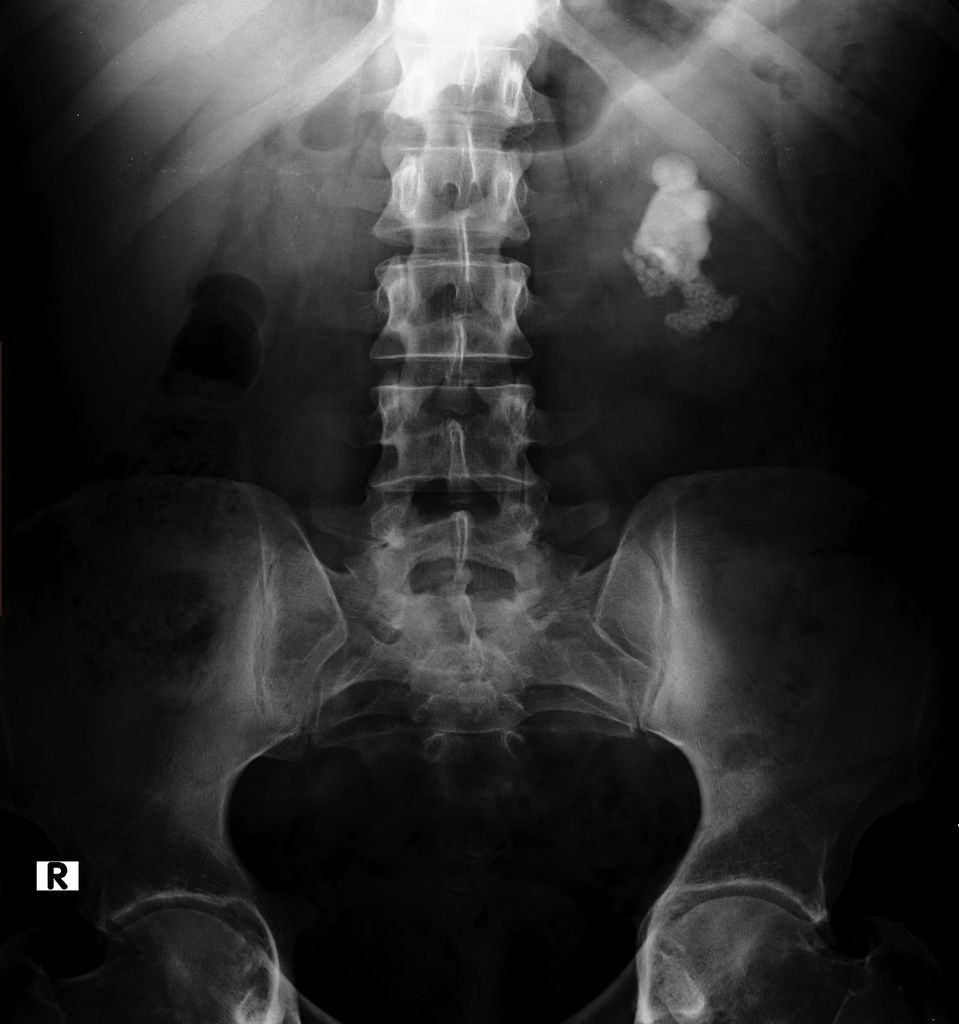
Kidney stones chest x ray. Uric acid stones and small kidney stones will not be revealed by a standard x ray. X ray examination on april 4 1924 showed the condition seen in figure 1 a much enlarged right kidney almost completely filled with overlapping masses of stones arranged in two groups that in the upper pole being somewhat smaller. They are thought to have lower attenuation than calcium stones and a slightly lower attenuation than struvite stones but there is overlap in.
Current guidelines state that kidney stone analysis should be performed using modern methods like infrared spectroscopy or x ray diffraction. When the stone is not on the focus the sound waves can damage the soft tissue of the kidney. Ct non contrast non contrast ct of the kidneys ureters and bladder is a quick and simple test that can be used to identify and locate calculi within the renal tract.
In this study an automated system is developed to detect kidney stones from x ray images. X ray vs ct scan kidney stones a 32 year old female asked. Although x rays are still sometimes used to evaluate kidney stones they are largely being replaced by ct scans which are a more accurate technique for imaging the urinary track and evaluating kidney stones.
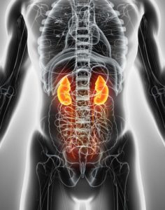
No stones were seen in the ureters or the left kidney. Doctors can use it to help them diagnose. A chest x ray uses very small amounts of radiation electromagnetic waves to create images of the structures inside your chest including your heart lungs airways and bones.
Cystine stones tend to be larger relative to other etiologies for stone disease and frequently coalesce to form large branching staghorn calculi. No stones uti etc. At mayo clinic we use fourier transform infrared spectroscopy ftir which is considered to be the reference method.
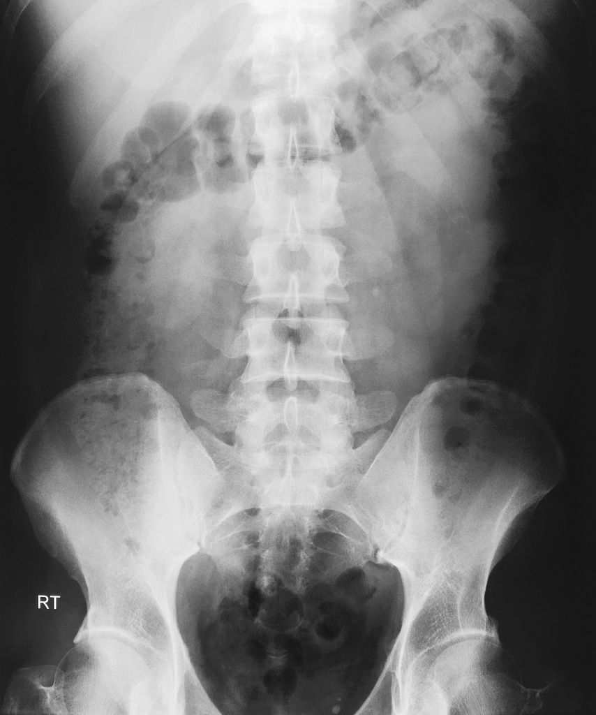
A kidney ureter and bladder kub study is an x ray study that allows your doctor to assess the organs of your urinary and gastrointestinal systems. This damage can be prevented by a feedback mechanism that determines the place of kidney stones depending on the images taken from eswl device. Low back pain would l spine xray have shown lower back tumor.
Ureteric Stone Radiology Case Radiopaedia Org

Xray Film Of A Patient With Multiple Kidney Stones In Both Kidneys Stock Photo Picture And Royalty Free Image Image 107589081

Artificial Kidney Ureters X Ray Kidney Kidney Health Medical Curiosities

Kidney Stone Renal Stone Renal Calculi Film X Ray Kub Stock Photo Picture And Royalty Free Image Image 47419529

The X Ray Abdomen Taken On 12 09 1997 Shows Gall Stones Staghorn Download Scientific Diagram

X Ray Kub Of A Patient With Bilateral Renal Stones Lar Open I
Staghorn Calculus Kidney Radiology Case Radiopaedia Org
Chest X Ray Kidney Cancer Cancer Research Uk

Xray Film Of A Patient With Multiple Kidney Stones Stock Photo Picture And Royalty Free Image Image 138777580

Welcome To Learningradiology Nephrocalcinosis Medullary Medullary Nephrocalcinosis Calculi Renal Stones Medullary Spo Radiology Radiology Imaging Renal

Pin By Kathy I On Radiology Radiography Radiology Medical

Case 205 Renal Stone Ileus Radiology

Xray Image Of Plain Kub Show Left Kidney Stone Stock Photo Picture And Royalty Free Image Image 40867964